Custom made, patient specific or standard model

When we talk about 3D printed medical models, it is important to distinguish between models representing custom-made medical devices, specific patients, and generic models. This distinction is important so that the choice of the model to be printed falls on the right solution, capable of responding effectively to the intended use and the appropriate regulatory framework.
We choose again to go into detail with the collaboration of Nils Winkes, Sales Manager Medical EMEA of Stratasys, whose task is precisely to enhance the skills of a medical public, towards the knowledge of additive technologies and 3D printing. We therefore thank Nils for the contribution through which this 2nd BIO3DNEWS was created and let’s get to the heart of the matter.
Medical 3D models
Custom and patient-specific, or personalized, medical 3D models are 3D printed from an STL file processed directly from diagnostic images (CT or MRI) and accurately respond to the specific morphological characteristics of the patient and his/her pathology. It is possible to obtain reproductions as faithful as possible to the real anatomy, in our case in polymer resin, which show the exact conformation not only of the limb, tissue or organ, but also of the clinical situation (malformation, contusion, fracture, cancerous mass, abrasion or other).
These 3D models are useful tools especially for preoperative practice, as they allow the surgeon to simulate the operation directly on the model, arriving in the room with a clear idea of how to conduct the operation in order to obtain an optimal result. Furthermore, these 3D printed models facilitate communication between doctor and patient, providing a practical demonstration of the operation to which the patient will be subjected and of the related pathological conditions of the same.
Stratasys 3D printers
For the production of 3D models, Stratasys DAP 3D printers can be used, with PolyJet technology, which guarantee excellent performance in terms of materials with realistic biomechanical characteristics, colors and resolution. These printing systems are present within the Bio3Dmodel competence center and are available both for direct purchase and for use through medical service. Among the materials that can be used we have:
- Agilus30: A photopolymer compatible with the Stratasys J850 3D printing system, ready to withstand long-term tearing, flexing and bending. Agilus30 is flexible and capable of offering a soft and realistic tactile response.
- Bone Matrix: the solution that allows you to create porous bone structures, fibrotic tissues and ligaments, reproducing the density and behavior of natural musculoskeletal parts.
- Tissue Matrix: soft but resistant, similar to human tissue, this material is used by Bio3DModel for 3D printing of soft and/or adipose tissue, fibrotic tissue, soft organs and tumor masses, with the possibility of testing on cutting, drilling and suturing.
- Radio Matrix: the radiopaque solution for 3D printing of VERO models and more, to give maximum realism to the parts to be subjected to diagnostics and medical imaging.
- Gel Matrix e linea Gel Support: the word itself says it, these are gel materials mainly used as support solutions for the creation of thin-walled and hollow-lumen vascular structures, but also suitable for the realistic printing of soft organs and adipose tissue.
There is also a specific range of VERO materials, rigid functional resins for the development of prototypes that can be used in the operating room (but not suitable for human contact).
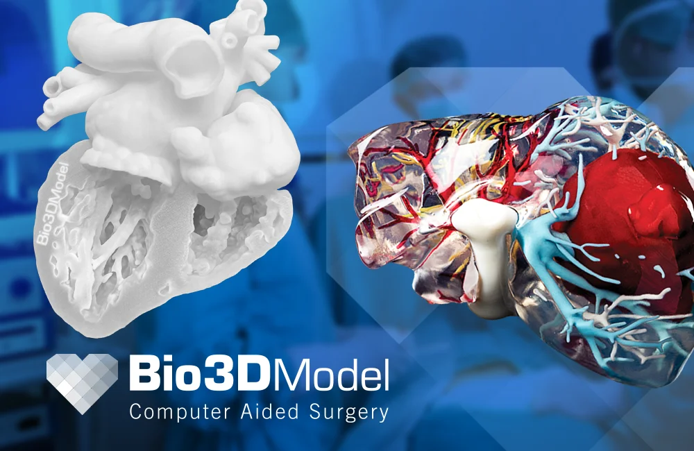
The use of colored resins, in particular, facilitates the differentiation of the areas to be treated, for example the right and left ventricle in the case of a 3D reproduction of the heart. The areas in question are divided already in the design phase through a segmentation process, which allows the delimitation of the different areas making them easily identifiable. This practice allows a better understanding of the situation, starting from the 3D model.
Going into the merits of medical 3D models, which from a regulatory point of view do not appear to be medical devices, it is useful to note how these 3D printed models lend themselves to being particularly useful allies for clinical training, facilitating the understanding of the anatomy of bones, tissues and organs, but also for the testing of new medical devices for post-operative recovery.
These are three-dimensional models capable of faithfully reproducing both the structure and the biomechanical behavior of the body part, however they cannot be considered as medical devices as they are not necessarily obtained from diagnostic images referring to a real patient but they can be modeled and segmented according to the training or testing needs.
Conclusion
In conclusion, in the context of medical training, these models allow for in-depth anatomical study of bones, organs and tissues.
We can therefore say that 3D printing, in the clinical field, continues to make progress in supporting training and surgical practice, with a view to obtaining better results in the surgical field.


