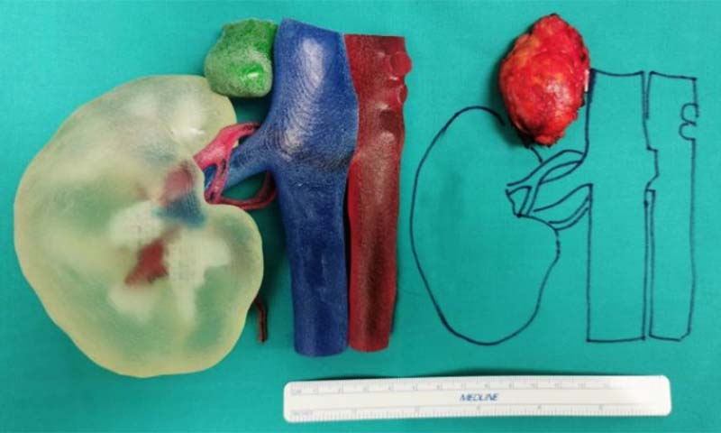
3D kidney model helps surgeons in laparoscopic adrenalectomy

3D kidney model helped surgeons at Santo Stefano hospital in Prato
The team of the General Surgery Unit of the Prato hospital, directed by Dr. Stefano Cantafio, within the Department of Surgical Specialties, directed by Dr. Stefano Michelagnoli, took advantage of this new frontier of precision surgery, applied for the first time in the Health Authority.
How was the 3D kidney model created?
The patient-specific model was 3D printed using Stratasys‘ proprietary J750 Digital Anatomy technology, reconstructed from diagnostic CT images, and represents a real-world case of a patient who underwent kidney surgery.
To print the 3D kidney model, transparent resin was used to allow internal visualization of the vessels and anatomical relationships. Furthermore, thanks to the possibility of printing with different colors, it was possible to make the tumor (printed in green) evident compared to the surrounding venous (in blue) and arterial (in magenta) vessels.
The 3D kidney model was made with colored and deformable resins that allowed the surgical simulation of the intervention in the preoperative phase. The blood vessels were reconstructed and printed to replicate the real vessels as faithfully as possible in both morphological and mechanical properties.
What was the 3D kidney model used for?
The 3D model was used to plan, study and correctly simulate the removal of the adrenal gland. With this model the surgeon was able to visualize and manipulate the vessels and the various anatomical relationships with the pathology itself.
Before the advent of 3D printing for the medical field, a complex surgical intervention was planned starting from two-dimensional black and white images produced by X-rays, CT and Magnetic Resonance Imaging. Thanks to this new technology, today it is possible to create a three-dimensional model of an organ or specific anatomical structures in real dimensions that can also simulate the morphological and mechanical properties of the organ itself, allowing to study the surgical intervention in advance.
Having the ability to plan the best strategic choice in advance, before intervening on the patient directly in the operating room, has allowed us to avoid intraoperative complications and reduce the risk of error.
“I am satisfied with the success of this delicate operation,” added Stefano Michelagnoli, director of the Department of Surgical Specialties. “This project is innovative, it allows us to obtain a personalized solution for each patient and to perform the operation safely by evaluating every aspect before arriving in the operating room, with advantages for both the patient and the professional. It has been applied for the first time at Santo Stefano and we plan to extend it to other surgical specialties.”
Stefano Michelagnoli, Director of the Department of Surgical Specialties at Santo Stefano Hospital in Prato


